ACG Clinical Guideline: Management of Patients With Acute Lo… : Official journal of the American College of Gastroenterology

INTRODUCTION
Acute overt lower gastrointestinal bleeding (LGIB) accounts for ˜20% of all cases of gastrointestinal (GI) bleeding, usually leads to hospital admission with invasive diagnostic evaluations, and consumes significant medical resources (1, 2, 3). Although most patients with acute LGIB stop bleeding spontaneously and have favorable outcomes, morbidity and mortality are increased in older patients and those with comorbid medical conditions (4).
An individual with acute LGIB classically presents with the sudden onset of hematochezia (maroon or red blood passed per rectum). However, in rare cases, patients with bleeding from the cecum/right colon can present with melena (black, tarry stools) (5). In addition, hematochezia can be seen in patients with brisk upper gastrointestinal bleeding (UGIB). Approximately 15% of patients with presumed LGIB are ultimately found to have an upper GI source for their bleeding (6). Historically, LGIB was defined as bleeding from a source distal to the Ligament of Treitz. However, bleeding from the small intestine (middle GI bleeding) is distinct from colonic bleeding in terms of presentation, management, and outcomes (7). For the purposes of this guideline, we define LGIB as the onset of hematochezia originating from either the colon or the rectum (8).
In this practice guideline, we discuss the main goals of management of patients with LGIB. First, we discuss the initial evaluation and management of patients with acute LGIB including hemodynamic resuscitation, risk stratification, and management of anticoagulant and antiplatelet agents (antithrombotic agents). We then discuss colonoscopy as a diagnostic and therapeutic tool including preparation, timing, and endoscopic hemostasis. Next, we outline non-colonoscopic diagnostic and therapeutic strategies for LGIB. Finally, we discuss prevention of recurrent LGIB and the role of repeat colonoscopy for recurrent bleeding events.
Each section of this document presents key recommendations followed by a summary of supporting evidence. A summary of the key recommendations is presented in Table 1.

Summary and strength of recommendations

Continued.
With the assistance of a health sciences librarian, a systematic search of the literature was conducted covering the years 1 January 1968 through 2 March 2015 in the PubMed and EMBASE databases and the Cochrane Library including the Cochrane Database of Systematic Reviews, the Database of Abstracts of Reviews of Effect, and Cochrane Central Register of Controlled Trials (CENTRAL). The PubMed search used a combination of Medical Subject Headings (MeSH), as well as terms appearing in titles and abstracts.
The strategy used to cover the lower gastrointestinal tract included (‘Exp Intestine, Large’[Mesh] OR ‘Exp Lower Gastrointestinal Tract’[Mesh] OR lower gastrointestinal[tiab] OR lower intestinal[tiab]). These terms were combined with terms for gastrointestinal bleeding including ‘Gastrointestinal Hemorrhage’[Mesh:noexp] OR rectal bleeding[tiab] OR colonic hemorrhage[tiab] OR colonic hemorrhages[tiab] OR colonic bleeding[tiab] OR hematochezia[tiab] OR haematochezia[tiab] OR rectal bleed[tiab] OR diverticular bleeding[tiab] OR diverticular bleed[tiab] OR diverticular hemorrhage[tiab] OR severe bleeding[tiab] OR active bleeding[tiab] OR melena[tiab] OR acute bleed[tiab] OR acute bleeding[tiab] OR acute haemorrhage[tiab] OR acute haemorrhage[tiab]) OR (LGIB[tiab] OR LIB[tiab]). The final group was limited to English language and human studies. Citations dealing with children and prostatic neoplasms were excluded. The following website will pull up the PubMed search strategy: http://tinyurl.com/ofnxphu.
Search strategies in EMBASE and the Cochrane Library databases replicated the terms, limits, and features used in the PubMed search strategy.
In addition to the literature search, we reviewed the references of identified articles for additional studies. We also performed targeted searches on topics for which there is relevant literature for UGIB but not LGIB including hemodynamic resuscitation/blood product transfusions and management of anticoagulant and antiplatelet medications.
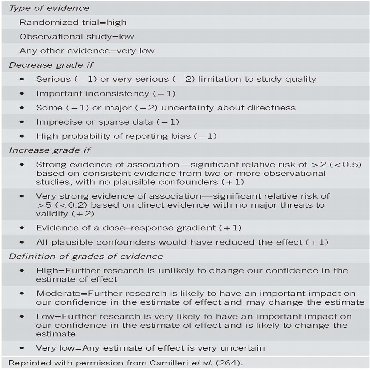
We used the GRADE system to grade the quality of evidence and rate the strength of each recommendation (9). The quality of evidence, which influences the strength of recommendation, ranges from “high” (further research is very unlikely to change our confidence in the estimate of effect) to “moderate” (further research is likely to have an important impact on our confidence in the estimate of effect and may change the estimate) to “low” (further research is very likely to have an important impact on our confidence in the estimate of effect and is likely to change the estimate) and “very low” (any estimate of effect is very uncertain). The strength of a recommendation is graded as strong when the desirable effects of an intervention clearly outweigh the undesirable effects and is graded as conditional when uncertainty exists about the trade-offs (9). Other factors affecting the strength of recommendation include variability in values and preferences of patients and whether an intervention represents a wise use of resources (9). In the GRADE system, randomized trials are considered high-quality evidence but can be downrated depending on the size, quality, and consistency of studies. Observational studies are generally rated as low-quality studies.
INITIAL ASSESSMENT
Evaluation and risk stratification
Recommendations
1.A focused history, physical examination, and laboratory evaluation should be obtained at the time of patient presentation to assess the severity of bleeding and its possible location and etiology. Initial patient assessment and hemodynamic resuscitation should be performed simultaneously (strong recommendation, very-low-quality evidence) (8, 10).
2.Hematochezia associated with hemodynamic instability may be indicative of an UGIB source, and an upper endoscopy should be performed. A nasogastric aspirate/lavage may be used to assess a possible upper GI source if suspicion of UGIB is moderate (strong recommendation, low-quality evidence) (6, 11, 12).
3.Risk assessment and stratification should be performed to help distinguish patients at high- and low-risk of adverse outcomes and assist in patient triage including the timing of colonoscopy and the level of care (conditional recommendation, low-quality evidence) (13, 14, 15, 16, 17, 18).
Summary of evidence
Initial assessment of the patient presenting with presumed acute LGIB should include a focused history, physical examination, and laboratory testing with the goal of determining the severity of bleeding, its possible location, and etiology (8, 10). The history obtained should include the nature and duration of bleeding and any associated symptoms that may suggest a specific source such as abdominal pain and diarrhea (colitis), and altered bowel habits and weight loss (malignancy). Likewise, past medical history elements should include any prior GI bleeding events, abdominal and/or vascular surgeries, peptic ulcer disease, inflammatory bowel disease, or abdominopelvic radiation therapy. It is also important to assess comorbidities including cardiopulmonary, renal, or hepatic disease that may put the patient at high risk of poor outcome and alter the management approach. Current or recent medication use should be noted, particularly those medications that may influence bleeding risk (nonsteroidal anti-inflammatory drugs (NSAIDs), antiplatelet agents, and anticoagulants). The physical examination should include the measurement of vital signs, including postural changes, to assess for hypovolemia. A cardiopulmonary, abdominal, and digital rectal examination should also be performed. The latter can detect potential anorectal bleeding sources and determine the color of the stool. Initial laboratory testing should include a complete blood count, serum electrolytes, coagulation studies, and a type and cross match.
Hematochezia associated with hemodynamic instability should lead to consideration of a brisk UGIB source, especially in at-risk patients such as those with a history of peptic ulcer disease or liver disease with portal hypertension and those using antiplatelet or anticoagulant medications (6, 11, 12, 19). An elevated blood urea nitrogen-to-creatinine ratio also suggests an UGIB source (likelihood ratio of UGIB with ratio >30:1 is 7.5) (10), whereas red blood and clots are unlikely to be from an upper gastrointestinal source (likelihood ratio 0.05) (10). If the likelihood of UGIB is high, an upper endoscopy should be performed. If suspicion for an UGIB source is modest, nasogastric aspirate/lavage can be used to assess possible UGIB (6, 11, 12). A positive nasogastric aspirate indicates a very high likelihood of an UGIB (likelihood ratio=11), whereas a negative aspirate makes an UGIB less likely but still possible (negative predictive value 64%, likelihood ratio=0.6) (20). Therefore, a positive or non-diagnostic (non-bloody, non-bilious) aspirate necessitates upper endoscopy before considering colonoscopy (12, 21). The nasogastric tube can be left in place to facilitate subsequent colon preparation (22).
Clinical data available at the time of initial patient evaluation can be used to identify patients at high risk for severe bleeding and other adverse outcomes. Several tools have been developed to assess risk in acute LGIB (Tables 2 and 3) (13, 14, 15, 16, 17, 18), although the number of available studies is modest in comparison with UGIB. Risk factors identified for poor outcome in LGIB include markers of hemodynamic instability at presentation (tachycardia, hypotension, and syncope), ongoing bleeding (gross blood on initial digital rectal examination and recurrent hematochezia), comorbid illnesses, age >60 years, a history of diverticulosis or angioectasia, an elevated creatinine, and anemia (initial hematocrit ≤35%). In general, the likelihood of an adverse outcome increases with the number of risk factors present (16). Monitoring in an intensive care setting should be considered in patients with high-risk features. These patients may also benefit from colonoscopy after a rapid bowel preparation or radiographic interventions.

Risk prediction tools for patients presenting with presumed LGIB

Risk factors for poor outcome in patients with LGIB
Hemodynamic resuscitation
Recommendations
4.Patients with hemodynamic instability and/or suspected ongoing bleeding should receive intravenous fluid resuscitation with the goal of normalization of blood pressure and heart rate before endoscopic evaluation/intervention (strong recommendation, very-low-quality evidence) (23, 24).
5.Packed red blood cells (RBCs) should be transfused to maintain the hemoglobin above 7 g/dl. A threshold of 9 g/dl should be considered in patients with massive bleeding, significant comorbid illness (especially cardiovascular ischemia), or a possible delay in receiving therapeutic interventions (conditional recommendation, low-quality evidence) (25, 26).
Summary of evidence
Patients with hemodynamic instability should receive intravenous fluid resuscitation (19, 23, 24). In UGIB, an intensive fluid (crystalloid) resuscitation strategy vs. standard of care may decrease mortality, myocardial infarction, and time in the intensive care unit. However, in the lone small study, these differences were not statistically significant (23, 24), and a specific resuscitation protocol was not outlined. In the critical care literature in general, there is considerable controversy regarding the timing, amount, and type of fluid resuscitation (27). However, there does not appear to be a benefit of colloid over crystalloid fluids (28). In addition, some patients will require blood transfusions. Transfusion strategies specific to LGIB have not been developed. Large observational studies and a meta-analysis of three small trials of UGIB suggest that blood transfusion compared with no transfusion is associated with an increased risk of rebleeding and possibly death (25, 29, 30, 31, 32). These findings are supported by results of a large randomized trial of patients with UGIB that found that a restrictive transfusion strategy with a transfusion threshold of hemoglobin <7 g/dl improved survival (95% vs. 91%) and decreased rebleeding (10% vs. 16%) when compared with a threshold of 9 g/dl (26). Patients with massive bleeding, acute coronary syndrome, symptomatic peripheral vascular disease, or a history of cerebrovascular disease were excluded, and all patients underwent upper endoscopy within 6 h of presentation. Therefore, patients with LGIB who have significant comorbid disease, massive, ongoing bleeding, or delayed therapeutic interventions may benefit from a more lenient blood transfusion threshold.
Management of coagulation defects
Recommendations
6.Endoscopic hemostasis may be considered in patients with an international normalized ratio (INR) of 1.5–2.5 before or concomitant with the administration of reversal agents. Reversal agents should be considered before endoscopy in patients with an INR >2.5 (conditional recommendation, very-low-quality evidence) (33, 34, 35).
7.Platelet transfusion should be considered to maintain a platelet count of 50 × 109/l in patients with severe bleeding and those requiring endoscopic hemostasis (conditional recommendation, very-low-quality evidence) (36, 37).
8.Platelet and plasma transfusions should be considered in patients who receive massive RBC transfusions (conditional recommendation, very-low-quality evidence) (37, 38, 39).
9.In patients on anticoagulant agents, a multidisciplinary approach (e.g., hematology, cardiology, neurology, and gastroenterology) should be used when deciding whether to discontinue medications or use reversal agents to balance the risk of ongoing bleeding with the risk of thromboembolic events (strong recommendation, very-low-quality evidence) (36, 40).
Summary of evidence
The management of anticoagulants and antiplatelet medications in the setting of LGIB requires consideration of the risk of ongoing bleeding and the risk of thromboembolic events and therefore requires an individualized approach. Observational studies of UGIB indicate that there is no increased risk of rebleeding following endoscopic hemostasis in patients with modest elevations in INR (1.5–2.7) (33, 34, 35, 41, 42, 43). A retrospective study of 98 patients with GI bleeding suggested that patients with an INR >4 had outcomes comparable to those with an INR in the 3–3.9 range, but these patients were not compared with patients with normal coagulation parameters (44). In addition, in these studies, the use and timing of reversal agents were difficult to discern. An INR >1.5 has been a predictor of mortality but not rebleeding in two large observational cohort studies presumably because INR is a strong indicator of underlying comorbid disease (33, 34). After adjustment for other potential confounders, the odds ratios for mortality in these studies were 1.96 (95% confidence interval (CI), 1.13–3.41) and 5.63 (95% CI, 3.09–10.27), respectively (30, 32). Careful attention should therefore be given to the management of comorbid illness in patients with coagulopathy.
Published standards in the hematology literature recommend platelet transfusion to maintain a platelet count of ≥50 × 109/l in patients with massive bleeding from any source (45, 46). There are no data to guide a threshold specific for gastrointestinal bleeding. Platelet transfusions should also be considered in patients who have a normal platelet count but receive massive RBC transfusions. Traditionally, massive transfusion has been defined as more than 10 units of packed RBCs within a 24-h period, but recent studies in the trauma literature define this threshold as 3 or more units of packed RBCs within 1 h (47). The trauma literature suggests a ratio of one unit of platelets and fresh frozen plasma per unit of RBCs transfused (38, 39, 48). A recent randomized trial indicated that a 1:1:1 ratio of plasma, platelets, and RBCs was associated with better hemostasis and fewer deaths due to exsanguination than a 1:1:2 protocol without a difference in other adverse events or death (37). The 1:1:1 ratio-based transfusion protocol likely applies outside the trauma setting(49), but no study has addressed a ratio-based transfusion protocol in gastrointestinal bleeding.
New target-specific oral anticoagulants including dabigatran, rivaroxaban, and apixaban are associated with an increased risk of GI bleeding. In a meta-analysis of 43 randomized controlled trials, the odds ratio for overall bleeding was 1.45 (95% CI, 1.07–1.97) (50). However, there is no direct evidence to guide the management of these agents in the setting of active GI bleeding. For elective procedures, a washout period based on the drug half-life is recommended (40) but may not be possible in patients with ongoing, acute bleeding or at high risk of thromboembolic events. In patients on target-specific oral anticoagulants, standard clotting tests may not reflect the degree of anticoagulation and thus cannot be used to guide the safety of endoscopic interventions. A reversal agent for dabigatran (idarucizumab) was recently approved by the Food and Drug Administration, and reversal agents for other non-vitamin K anticoagulants are in development (51). However, these antidotes may increase the risk of thrombosis (36, 40).
Therefore, a multidisciplinary approach involving hematology, cardiology/neurology, and gastroenterology is necessary when managing patients on anticoagulant medications, particularly if newer target-specific oral agents are involved to optimally balance the risk of ongoing bleeding with the risk of thromboembolic events. Please see the section on recurrent bleeding for recommendations regarding aspirin and antiplatelet medications.
COLONOSCOPY
Colonoscopy as a diagnostic tool
Recommendations
10. Colonoscopy should be the initial diagnostic procedure for nearly all patients presenting with acute LGIB (strong recommendation, low-quality evidence) (52).
11. The colonic mucosa should be carefully inspected during both colonoscope insertion and withdrawal, with aggressive attempts made to wash residual stool and blood in order to identify the bleeding site (53). The endoscopist should also intubate the terminal ileum to rule out proximal blood suggestive of a small bowel lesion (conditional recommendation, very-low-quality evidence).
Summary of evidence
Colonoscopy has both diagnostic and therapeutic roles in acute LGIB. The goal of colonoscopy in LGIB is to identify the site of bleeding and perform hemostasis, if indicated. The diagnostic yield of colonoscopy in this patient population ranges from 48 to 90% (52, 54). The most common causes of acute severe LGIB include diverticulosis, angioectasia, post-polypectomy bleeding, and ischemic colitis. Other less common causes include colorectal polyps/neoplasms, Dieulafoy’s lesions, inflammatory bowel disease, and anorectal conditions including solitary rectal ulcer, radiation proctitis, and rectal varices (55, 56). It is imperative to carefully inspect the colonic mucosa both on insertion and withdrawal, as culprit lesions often bleed intermittently and may be missed when not actively bleeding. The endoscopist should intubate the terminal ileum to rule out proximal blood suggestive of a small bowel lesion. An adult or pediatric colonoscope with a large working channel (at least 3.3 mm) should be used because the larger working channel facilitates suctioning of blood, clots, and residual stool, and allows for the passage of large diameter (e.g., 10 Fr) endoscopic hemostasis tools. In addition, the use of a water-jet irrigation device (foot pedal controlled by the endoscopist) is recommended to facilitate removal of adherent material and residue from the colonic mucosa.
Bowel preparation
Recommendations
12. Once the patient is hemodynamically stable, colonoscopy should be performed after adequate colon cleansing. Four to six liters of a polyethylene glycol-based solution or the equivalent should be administered over 3–4 h until the rectal effluent is clear of blood and stool. Unprepped colonoscopy/sigmoidoscopy is not recommended (strong recommendation, low-quality evidence) (10, 11, 19).
13. A nasogastric tube can be considered to facilitate colon preparation in high-risk patients with ongoing bleeding who are intolerant to oral intake and are at low risk of aspiration (conditional recommendation, low-quality evidence) (8, 57).
Summary of evidence
Colonoscopy should be performed after adequate preparation (11, 12, 22, 58). Preparation of the colon facilitates endoscopic visualization and diagnosis, and may reduce the risk of bowel perforation. Although there have been no head-to-head comparisons, studies using large volume (4–6 l), rapid (3–4 h) purge protocols using polyethylene glycol-based solutions with colonoscopy performed within 1–2 h of preparation completion report high rates of definitive diagnosis (22–42%) and hemostasis (34%) (11, 12, 22). Lower volume or alternative colon preparation solutions have been evaluated in the setting of colorectal cancer screening and surveillance but not in the setting of LGIB (59). Regardless of the solution used, it is important to clear the colon of stool, clots, and old blood to facilitate visualization and localization of the bleeding source. Many patients with acute LGIB are unable to tolerate rapid colon preparation and thus a nasogastric tube can be placed to facilitate this process (11, 22). In studies of urgent colonoscopy, as many as one-third of patients required a nasogastric tube to facilitate rapid bowel preparation (22). In addition, administration of a prokinetic/antiemetic agent immediately before initiating the colon preparation may reduce nausea and facilitate gastric emptying (8, 57). Complications of colon preparation with polyethylene glycol are rare but include aspiration pneumonia, as well as fluid and electrolyte abnormalities (12, 60). Aspiration precautions should be used particularly in older and debilitated patients.
Unprepped sigmoidoscopy/colonoscopy in the setting of acute LGIB is not recommended. In studies of urgent colonoscopy without oral or rectal preparation, cecal intubation rates are low (55–70%) (61, 62, 63). Recent prospective pilot data in severe LGIB subjects (n=12) reported the feasibility and safety of “unprepared hydroflush colonoscopy” that combined three 1-liter tap water enemas, a water-jet pump irrigation system, and a mechanical suction device to cleanse the colon (64). However, localization of bleeding, in particular diverticular bleeding, can be difficult in the setting of residual blood and stool, and poor visualization may also increase the risk of perforation. Therefore, this method is recommended only as an adjunct to appropriate oral preparation until further data are available.
Timing of colonoscopy
Recommendations
14. In patients with high-risk clinical features and signs or symptoms of ongoing bleeding, a rapid bowel purge should be initiated following hemodynamic resuscitation, and a colonoscopy performed within 24 h of patient presentation after adequate colon preparation to potentially improve diagnostic and therapeutic yield (conditional recommendation, low-quality evidence) (11, 22).
15. In patients without high-risk clinical features or serious comorbid disease or those with high-risk clinical features without signs or symptoms of ongoing bleeding, colonoscopy should be performed next available after a colon purge (conditional recommendation, low-quality evidence) (52, 65).
Summary of evidence
Studies of timing of colonoscopy in the setting of acute LGIB are limited. Table 4 summarizes the three existing prospective studies of urgent colonoscopy for acute LGIB. In a prospective study of 48 patients with severe diverticular bleeding who underwent colonoscopy within 12 h with endoscopic hemostasis, and 73 historical controls who underwent colonoscopy within 12 h without endoscopic therapy, outcomes were significantly better in the endoscopic hemostasis group: rebleeding (0% vs. 53%); emergency surgery (0% vs. 35%); and hospital length of stay (median 2 days vs. 5 days) (22). In addition, untreated stigmata of hemorrhage were predictive of subsequent outcomes in this study and a subsequent larger series, although the overall number of cases in each category is small and therefore the estimates may be imprecise. Rebleeding was seen in 84% of patients with active bleeding at endoscopy (n=16/19), 60% of patients with a non-bleeding visible vessel (n=3/5), and 43% with adherent clot (n=6/14) (22, 66). A trial of 100 patients with acute LGIB randomized to colonoscopy within 8 h of presentation or standard of care (colonoscopy next available or if unstable nuclear scintigraphy and angiography) found that urgent interventions significantly improved definitive diagnoses (42% vs. 22%, odds ratio, 2.6; 95% CI, 1.1–6.2) but not rebleeding, surgery, or length of stay (11). No stigmata were identified on elective colonoscopy, and the therapeutic yield was higher but not statistically significantly different in the urgent vs. elective group (34% endoscopic therapy vs. 20% angiographic therapy). In another trial of 72 patients randomized to colonoscopy within 12 h or delayed colonoscopy (30–60 h), there were no differences in rebleeding, diagnoses, or the need for therapy between the groups (6). Overall, retrospective studies support that urgent colonoscopy (defined variably as colonoscopy within 12–24 h) improves diagnostic and therapeutic yield (52). In addition, studies have found that earlier time to colonoscopy is associated with reduced hospital length of stay likely because of more efficient discharge after a negative exam (52, 65, 67). It is not clear whether urgent colonoscopy improves important clinical outcomes such as rebleeding and the need for surgery. However, because diagnostic yield is improved with earlier timing, the lack of a significant benefit in existing studies may reflect inadequate statistical power or insufficient endoscopic therapy.

Prospective studies of urgent colonoscopy for acute LGIB
Endoscopic hemostasis therapy
Recommendations
16. Endoscopic therapy should be provided to patients with high-risk endoscopic stigmata of bleeding: active bleeding (spurting and oozing); non-bleeding visible vessel; or adherent clot (strong recommendation, low-quality evidence) (22).
17. Diverticular bleeding: through-the-scope endoscopic clips are recommended as clips may be safer in the colon than contact thermal therapy and are generally easier to perform than band ligation particularly for right-sided colon lesions (conditional recommendation, low-quality evidence) (68, 69).
18. Angioectasia bleeding: noncontact thermal therapy using argon plasma coagulation is recommended (conditional recommendation, low-quality evidence) (75, 76).
19. Post-polypectomy bleeding: mechanical (clip) or contact thermal therapy, with or without the combined use of dilute epinephrine injection, is recommended (strong recommendation, very-low-quality evidence) (70, 71).
20. Epinephrine injection therapy (1:10,000 or 1:20,000 dilution with saline) can be used to gain initial control of an active bleeding lesion and improve visualization but should be used in combination with a second hemostasis modality including mechanical or contact thermal therapy to achieve definitive hemostasis (strong recommendation, very-low-quality evidence) (11, 22, 52).
Summary of evidence
Colonoscopy with endoscopic hemostasis for colonic bleeding is safe. Adverse events were reported in 0.3–1.3% of more than 2,400 colonoscopies performed for acute LGIB (69, 72). Moreover, endoscopic hemostasis in the colon appears to be effective, although the optimal technique has not yet been fully characterized. Endotherapy options for acute LGIB include injection (most commonly dilute epinephrine), contact thermal therapies (bipolar/multipolar electrocoagulation, heat probe), noncontact thermal therapy (argon plasma coagulation), through-the-scope clipping devices, and band ligation. Emerging endoscopic treatments include hemostatic topical sprays/powders and large-sized over-the-scope clipping devices (73, 74). Each of these therapeutic modalities, used as monotherapy or in combination, has been reported to be safe and effective in controlling bleeding. In contrast to the numerous randomized comparative studies and meta-analyses evaluating endoscopic hemostasis modalities in acute UGIB, there have been no such studies in acute LGIB. Endoscopic treatments have most commonly been reported as individual case reports, retrospective cohort studies, or prospective, non-randomized case series with small numbers of patients. Thus, the endoscopic hemostasis modality selected by the endoscopist is generally guided by the source of bleeding, access to the bleeding site, and experience with the various hemostasis device options.
The most common causes of LGIB amenable to endotherapy are diverticulosis, angioectasia, and post-polypectomy bleeding (56). Endoscopic therapy for each of these bleeding etiologies will be discussed below.
Diverticular hemorrhage
Diverticular bleeding is arterial, typically presents as painless hematochezia, and usually occurs from either the neck or the dome of the diverticulum (22). Patients with diverticular bleeding are candidates for endoscopic treatment if active bleeding (spurting or oozing), a non-bleeding visible vessel, or an adherent clot (that cannot be removed with vigorous washing and suctioning) is found at the time of colonoscopy (22). As noted above, these stigmata of hemorrhage predict a high risk of rebleeding without treatment (66).
Jensen et al. reported a prospective case series of 10 patients presenting with severe hematochezia found to be from a definitive diverticular source at the time of urgent colonoscopy. Endoscopic treatments included injection of dilute epinephrine (1:20,000 admixture with saline, in 1 or 2 ml aliquots per injection in four quadrants), as monotherapy for patients with active bleeding (n=5), and bipolar thermal coagulation (using 10–15 W with moderate appositional pressure applied in 1-s intervals until vessel flattening was achieved) for those with a non-bleeding visible vessel (n=2). For patients with an adherent clot (n=3), dilute epinephrine was injected circumferentially around the site of bleeding, the clot was removed using a colon polyp snare, and any underlying stigmata were treated with bipolar thermal coagulation as described above (22). None of the 10 patients treated endoscopically had recurrent bleeding or required surgery. In a pooled analysis of case series (including 847 patients) evaluating colonoscopy and endoscopic hemostasis for diverticular bleeding, Strate et al. (69) reported that following endoscopic hemostasis (n=137), early rebleeding occurred in 8% and late rebleeding in 12% of patients. There was no apparent advantage to combined endoscopic hemostasis over monotherapy.
Endoscopic clips are an attractive treatment modality for diverticular bleeding. Compared with contact thermal therapies, clips avoid the theoretical risk of transmural injury and perforation in the thin-walled colon. In addition, improved clip design including greater tensile strength and the ability to rotate and open/close the clip before deployment has made clips easier to use for bleeding control (75, 76, 77, 78, 79). Control of diverticular bleeding using clips can be accomplished either by targeted clip placement directly on the bleeding stigma or by closure of the diverticular orifice in a “zipper-like” manner resulting in bleeding tamponade (79). When active bleeding is present, dilute epinephrine (0.5–2 ml per injection) can be injected in or around the diverticulum to slow bleeding, improve visibility, and facilitate clip placement (68). In the setting of a small or deep bleeding diverticulum, a translucent cap can be placed onto the tip of the colonoscope, enabling eversion of the diverticulum for more precise localization and treatment of the bleeding lesion (68). Moreover, injection can also be used to evert the dome of the diverticulum and improve access to the bleeding site followed by clip placement (8).
In the aforementioned pooled analysis by Strate and Naumann, no early rebleeding was reported after endoscopic clipping of diverticular bleeding; however, late rebleeding occurred in 17% (8). More recently, in a retrospective case series from two Veterans Affairs hospitals, Kaltenbach reported on the short- and long-term outcomes of endoscopic clipping in 24 patients with definitive diverticular hemorrhage (68). Successful endoscopic hemostasis was achieved in 21 (88%) using clips as monotherapy or in combination with epinephrine injection in the setting of active bleeding. There was no early rebleeding or adverse events (e.g., perforations). Late rebleeding (≥30 days following initial endoscopic hemostasis) occurred in 24%. Of the three patients in whom primary hemostasis was not achieved, two required emergency hemicolectomy and one patient received angiographic embolization.
Case series including a total of 36 patients report good safety and efficacy of endoscopic band ligation for the treatment of diverticular bleeding with stigmata of recent hemorrhage (80, 81, 82). The banding technique described includes identification of the culprit diverticulum, marking of the site with a clip or India ink, followed by withdrawal of the colonoscope. A band ligation device is then loaded onto a gastroscope (if the bleeding lesion is located in the left colon) or a pediatric colonoscope. Once the lesion is re-identified, it is suctioned into the banding device, and the band is deployed as is done in the treatment of variceal hemorrhage. Recently, Shibata et al. (83) reported on 27 cases of definitive colonic diverticular hemorrhage effectively treated (hemostasis achieved in 96.3%) using band ligation in combination with a disposable, transparent soft hood attached to the tip of the colonoscope. The hood allows improved visualization of diverticula and exposure of high-risk stigmata. Caution, however, should be exercised when contemplating using band ligation for a right side colonic diverticular bleed. Ex vivo colon specimen data have demonstrated serosal entrapment and inclusion of the muscularis propia post band ligation in the right colon (84, 85). The left colon, likely due to its thicker mucosal wall, had limited submucosal involvement and only a single site of muscularis propria involvement (84).
The use of Doppler ultrasound probe monitoring has been reported as an adjunct to endoscopic treatment. In a study of 46 patients with diverticular bleeding, 24 were found to have major stigmata of hemorrhage at the time of colonoscopy (66). Doppler ultrasound probe noted arterial flow in 92% (and no flow in those without major stigmata). After treatment, no patient had residual blood flow and no patient experienced rebleeding at 30 days. However, there was no comparison with patients undergoing endoscopic treatment without Doppler probe guidance. Therefore, Doppler ultrasound probe guidance holds promise for improving the effectiveness of endoscopic hemostasis in diverticular bleeding, but further data are needed.
After endoscopic treatment, an India ink tattoo or clip (if not already used for hemostasis) should be placed adjacent to the culprit lesion to assist in re-localization should rebleeding occur (8, 82).
Angioectasia
Angioectasias are common in the right colon and in the elderly (86, 87). Colonic angioectasias, including radiation proctopathy, usually present with occult bleeding but can present with overt hematochezia, especially in patients using anticoagulant/antiplatelet therapy (8, 57). Endoscopic hemostasis therapy is indicated if there is evidence of acute or chronic blood loss (88). Contact and noncontact thermal endoscopic therapies are effective for treatment of angiodyplasia. Noncontact thermal therapy (argon plasma coagulation) is more commonly used because it is easy to use, safe, efficient, and has been shown to improve hemoglobin levels and reduce the frequency of blood transfusions (89, 90). Typical argon plasma coagulation power settings for the treatment of colonic angioectasia are 20–60 W (lower power used in the right colon) with an argon gas flow rate 1–2.5 l/min (89, 90). Lesions are obliterated using focal pulses of 0.5–2-s duration. Larger angioectasia (>10 mm) and those located in the right colon may be lifted using submucosal saline injection before coagulation (89, 91).
Post-polypectomy bleeding
Post-polypectomy bleeding can occur immediately or days to weeks following polyp removal (92). Risk factors for post-polypectomy bleeding include large polyp size (>2 cm), thick stalk, right colon location, and resumption of antithrombotic therapy. Endoscopic hemostasis treatments for post-polypectomy bleeding include endoscopic clipping, thermal contact, with or without the combined use of dilute epinephrine injection, and band ligation. Use of through-the-scope clipping, with or without epinephrine injection, may be preferred in order to limit additional tissue injury that occurs with contact thermal coagulation therapy (92).
Hemostatic topical powders/sprays have recently been reported as an endotherapy options for acute LGIB (93). These powders/sprays (Hemostatic Agent TC-325 (Hemospray, Cook Medical, Winston-Salem, NC), EndoClot polysaccharide hemostatic system (EndoClot Plus Inc., Santa Clara, CA), and Ankaferd Bloodstopper (Ankaferd ilac kozmetik A.S., Istanbul, Turkey)) are delivered through the working channel of the endoscope and are intended to control “actively” bleeding lesions. There are a limited number of case reports and small case series reporting on these modalities as primary or salvage therapy in post-polypectomy bleeding, colonic ulcerations including solitary rectal ulcer, radiation proctitis, colorectal neoplasia, and portal hypertensive colopathy (94, 95, 96, 97, 98). In addition, an over-the-scope clip (OTSC, Ovesco Endoscopy, Tubingen, Germany), made from a nitinol alloy, has been applied as salvage therapy in post-polypectomy bleeding (99). This clipping device is loaded onto an endoscope and deployed in a similar manner as a band-ligating device.
Acute LGIB etiologies such as ischemic colitis, colitis due to inflammatory bowel disease, and colorectal neoplasms are generally not amenable to durable endoscopic hemostasis and are treated with supportive medical and/or surgical care of the underlying etiology.
Role of repeat colonoscopy in the setting of early recurrent bleeding
Recommendations
21. Repeat colonoscopy, with endoscopic hemostasis if indicated, should be considered for patients with evidence of recurrent bleeding (strong recommendation, very-low-quality evidence) (68, 79).
Summary of evidence
The rate of rebleeding in patients with acute LGIB is poorly characterized. In randomized controlled studies, early rebleeding (defined as rebleeding prior to hospital discharge) following urgent colonoscopy is reported to be 22% and late rebleeding (defined as rebleeding after hospital discharge) is 16% (6, 11). Factors that may contribute to early or late rebleeding include underlying comorbid conditions, concurrent medication use (e.g., NSAIDs, antiplatelet agents, anticoagulants), source of index bleeding, and initial hemostasis modality (100). There are no published studies that directly evaluate the role of repeat colonoscopy in patients with early or late recurrent LGIB. However, small case series indicate that the yield of repeat colonoscopy for early rebleeding from a diverticular source is fairly high (20%) (79). In this setting, the patient often remains in the hospital with a recently prepped colon, and repeat colonoscopy can be performed promptly.
NON-COLONOSCOPY INTERVENTIONS
Recommendations
22. A surgical consultation should be requested in patients with high-risk clinical features and ongoing bleeding. In general, surgery for acute LGIB should be considered after other therapeutic options have failed and should take into consideration the extent and success of prior bleeding control measures, severity and source of bleeding, and the level of comorbid disease. It is important to very carefully localize the source of bleeding whenever possible before surgical resection to avoid continued or recurrent bleeding from an unresected culprit lesion (conditional recommendation, very-low-quality evidence).
23. Radiographic interventions should be considered in patients with high-risk clinical features and ongoing bleeding who have a negative upper endoscopy and do not respond adequately to hemodynamic resuscitation efforts and are therefore unlikely to tolerate bowel preparation and urgent colonoscopy (strong recommendation, very-low-quality evidence) (101, 102).
24. If a diagnostic test is desired for localization of the bleeding site before angiography, computed tomographic (CT) angiography should be considered (conditional recommendation, very-low-quality evidence) (69).
Summary of evidence
A number of radiographic modalities can be utilized in the setting of presumed acute LGIB. Few studies have compared radiographic interventions with colonoscopy. In one randomized trial evaluating colonoscopy within 8 h of admission compared with elective colonoscopy if hemodynamically stable or tagged RBC scan followed by angiography if ongoing bleeding, more diagnoses and therapeutic interventions were made in the urgent colonoscopy arm (11). Retrospective studies also suggest the superior diagnostic and therapeutic yield of colonoscopy over radiographic algorithms (101, 102). In contrast to radiographic modalities, colonoscopy can provide a definitive diagnosis and treatment in the absence of active bleeding at the time of the exam. Nonetheless, in some patients brisk, ongoing hematochezia precludes adequate hemodynamic resuscitation and bowel preparation before colonoscopy. In this small subset, angiography can provide both localization and treatment. Angiography localizes a LGIB source in 25–70% of exams (103, 104). A systematic review found that super-selective angiographic embolization achieves immediate hemostasis in 40–100% of cases of diverticular bleeding with a rebleeding rate ranging from 0 to 50% (105). Bowel ischemia is reported in as many as one-third of patients following super-selective embolization (105), although the rate of ischemia is lower (1–4%) in more recent series (103, 106). Because angiography relies on active bleeding and has the potential for serious complications, it should be reserved for patients with very brisk, ongoing bleeding.
There is a considerable debate regarding the utility of tagged RBC scintigraphy to localize GI bleeding before angiography. Some retrospective case series suggest that a screening-tagged RBC scintigraphy study increases the diagnostic yield of angiography and enables targeted contrast injection (107, 108, 109). Other series have found that the diagnostic yield of angiography is similar with or without a preceding tagged RBC scintigraphy (110, 111). If tagged RBC scintigraphy is positive, angiography should be performed immediately following to maximize the chance of a positive test. The ability of tagged RBC scintigraphy to accurately localize a bleeding source is suboptimal (65–80%) (69, 72, 112), and bleeding location should be confirmed before surgical resection particularly if the tagged RBC scintigraphy is positive only on delayed images (113, 114). One advantage of tagged RBC scintigraphy is the ability to perform repeated scans after the initial injection of tagged cells. This makes RBC scintigraphy most suitable for the evaluation of intermittent, obscure-overt GI bleeding (107, 115).
CT angiography or multi-detector row CT scan is another diagnostic modality for GI bleeding that is widely available and highly accurate at localizing the bleeding site (nearly 100%) (69). However, in the only back-to-back comparison, tagged RBC scintigraphy was positive in 46% of patients and CT angiography in 27% of patients (111). Nonetheless, only 2 of 11 patients with a positive RBC scintigraphy and negative CT angiography went on to have bleeding requiring treatment. Therefore, although tagged RBC scintigraphy may be more sensitive for bleeding, CT angiography is a reasonable first-line screening test if needed before angiography or emergent surgery because it is more expedient and accurate than tagged RBC scintigraphy. Standard precautions should be taken to avoid contrast-induced nephropathy, particularly as patients may undergo subsequent angiography with administration of arterial contrast (116).
A surgery consultation should be requested in patients with brisk, ongoing LGIB. The quality of the evidence regarding surgery for acute LGIB is poor and mostly derived from small, retrospective reviews. Some studies report high overall mortality (up to 27%) after emergent total abdominal colectomy for massive LGIB (117), whereas others note no difference in morbidity or mortality when comparing limited resection with total colectomy for bleeding (118). Not surprisingly, the rebleeding rate is higher in patients after limited resection than total colectomy (18% vs. 4% in one study of 77 patients) (118). In general, surgery for acute LGIB should be considered only after other therapeutic options have failed and should take into consideration the extent and success of prior bleeding control measures, severity and source of bleeding, and the level of comorbid disease. It is important to very carefully localize the source of bleeding whenever possible before surgical resection to avoid continued or rebleeding from an unresected culprit lesion.
PREVENTION OF RECURRENT LOWER GI BLEEDING
Recommendations
25. Non-aspirin NSAID use should be avoided in patients with a history of acute LGIB particularly if secondary to diverticulosis or angioectasia (strong recommendation, low-quality evidence) (119, 120, 121).
26. In patients with established high-risk cardiovascular disease and a history of LGIB, aspirin used for secondary prevention should not be discontinued. Aspirin for primary prevention of cardiovascular events should be avoided in most patients with LGIB (strong recommendation, low-quality evidence) (122, 123, 124).
27. In patients on dual antiplatelet therapy or monotherapy with non-aspirin antiplatelet agents (thienopyridine), non-aspirin antiplatelet therapy should be resumed as soon as possible and at least within 7 days based on multidisciplinary assessment of cardiovascular and GI risk and the adequacy of endoscopic therapy (as above, aspirin use should not be discontinued). However, dual antiplatelet therapy should not be discontinued in patients with an acute coronary syndrome within the past 90 days or coronary stenting within the past 30 days. (strong recommendation, low-quality evidence) (122, 125, 126).
Summary of evidence
Patients with bleeding from colonic diverticula or angioectasia are prone to recurrent bleeding events. The rate of diverticular hemorrhage recurrence at 1 year in patients who do not undergo surgical treatment was reported at 9% in a population-based study (3) but was considerably higher (47%) in a single-center study of patients with definitive diverticular bleeding (127). It is not clear that endoscopic therapy of diverticular stigmata decreases the rate of recurrent bleeding, particularly because bleeding may arise from any existing diverticulum. Rates of late rebleeding are reported in ˜15% of patients after combination injection plus thermal or clip therapy, with variable follow-up periods (69).
Angioectasias are also prone to rebleeding, and new lesions may form throughout the GI tract. In a systematic review, the rate of rebleeding with conservative/placebo therapy ranged from 37 to 45% at 1 year and 58 to 64% at 2 years (128). The authors rated the evidence for treatment with thalidomide or estrogen plus progesterone as low and that for octreotide as insufficient. Medical therapies resulted in a higher rate of complications than placebo. Small, retrospective studies have examined the use of argon plasma coagulation, heater probe, and monopolar thermal coagulation in the treatment of angioectasia. Rates of rebleeding were no different between any of the endoscopic modalities and conservative care (128).
Risk factors for recurrent LGIB are not well-studied. In one study of 83 patients with incident diverticular bleeding events who were followed for an average of 34 months, no predictors were identified including age, gender, blood transfusion requirements, hospital length of stay, endoscopic stigmata, or a previous history of bleeding (3). However, risk factors for incident diverticular bleeding events include obesity, physical inactivity, hypertension, hyperlipidemia, and chronic renal insufficiency (129, 130, 131, 132). It is not known whether modification of these risk factors reduces the risk of recurrent events.
Several studies indicate that NSAIDs increase the risk of both incident and recurrent LGIB. A prospective study of 132 patients hospitalized with diverticular bleeding found that recurrence was 77% among patients who continued NSAID use vs. 9% in those who discontinued (121). In another study of 342 patients with LGIB (50% due to a diverticular source) with a mean follow-up of 19 months, the cumulative rebleeding rate was 17% in patients on no antiplatelet medications, 31% on monotherapy, and 47% on dual antiplatelet therapy (120). In a multivariate analysis, the relative risk for NSAID use was 2.0 (95% CI, 1.2–3.3), for non-aspirin antiplatelet drugs 1.8 (95% CI, 1.0–2.3), and for low-dose aspirin 1.3 (95% CI, 0.8–2.3). The risk was higher in users of dual therapy than monotherapy (relative risk, 1.8; 95% CI, 1.0–3.2). On the basis of this evidence, non-aspirin NSAID use should be avoided in patients with a history of acute LGIB, particularly if secondary to a diverticular source. Although COX-2 selective agents are associated with a lower risk of UGIB than non-selective agents, their safety in LGIB is less clear as the results of studies are mixed perhaps due to the relative antiplatelet effect of different formulations or concomitant low-dose aspirin use in some studies (133, 134, 135).
The risk of antiplatelet-associated rebleeding events may be higher in LGIB than UGIB given the lack of prophylactic measures including proton pump inhibitor (PPI) therapy and Helicobacter pylori treatment. In a study of aspirin, clopidogrel, and PPI therapy following percutaneous coronary intervention, LGIB was more common than UGIB (74% vs. 26% of bleeds) (136). Similarly, a large retrospective study of US Veterans found that the incidence of lower GI events in patients on complex antithrombotic therapy was higher than that of upper GI events (70 vs. 20/1,000 patient-years) (137). In addition, the likelihood of early and late rebleeding in the setting of aspirin is likely to vary according to bleeding etiology and adequacy and type of initial hemostasis (for early rebleeding). As noted above, long-term recurrence is common in patients with bleeding from angioectasia and diverticulosis. The risk of early rebleeding in the setting of antiplatelet or anticoagulant use may be higher with thermal contact hemostasis methods than with mechanical methods (clips) (138).
The available data on resumption of aspirin in the setting of GIB are from patients with bleeding peptic ulcers. In a randomized controlled trial of immediate resumption of low-dose aspirin plus PPI vs. placebo plus PPI after endoscopic control of ulcer bleeding, there was no significant difference in rebleeding (10% vs. 5%). However, 60-day all-cause mortality (1% vs. 13%), as well as mortality secondary to cardiovascular, cerebrovascular, or gastrointestinal complications, was significantly lower in patients treated with aspirin (123). In a hospital-based cohort study, the risk of death was sixfold higher in patients with peptic ulcer bleeding who stopped aspirin vs. those who did not (124). Data from patients undergoing polypectomy suggest that the risk of bleeding is similar in patients discontinuing vs. continuing aspirin (139). Therefore, aspirin for secondary prophylaxis in patients with established cardiovascular disease should not be discontinued in the setting of LGIB to avoid thromboembolic events. In contrast, in patients without established cardiovascular disease and who are not at high risk for cardiovascular events, aspirin (as primary prophylaxis) has been shown to have little net benefit (0.07% absolute risk reduction per year) (140), and should be avoided in the setting of LGIB.
The decision to use other antiplatelet and anticoagulant medications after an episode of LGIB requires a multidisciplinary approach that takes into consideration the risk of bleeding, as well as the risk of thromboembolic events (138). During the first 30 days following coronary stenting, the risk of death and myocardial infarction is doubled in patients who discontinue clopidogrel (126). The risk associated with discontinuation is also high in the first 90 days following an acute coronary syndrome. However, discontinuation for up to 7 days in patients with more distant coronary stenting or coronary syndrome appears to be safe as long as aspirin therapy is continued.
CONCLUSION
In this guideline, we sought to evaluate and summarize the literature on major issues in the management of patients with acute LGIB. In general, we found the quality of the existing evidence to be low. There are only a few small, randomized trials of patients with acute LGIB, and therefore we relied heavily on case–control or cohort studies, case series, systematic reviews, or indirect evidence from trials of UGIB. Despite these limitations, we strongly endorse some of the recommendations because the potential benefits appear to outweigh the risk of harm. An approach to patients presenting with acute LGIB is outlined in Figure 1. To summarize, patients presenting with acute severe hematochezia should undergo a focused evaluation simultaneous with hemodynamic resuscitation. An upper GI bleeding source needs to be excluded in patients with hematochezia and hemodynamic instability. Colonoscopy following a colon purge is the initial test of choice in most patients presenting with acute hematochezia. In patients with high-risk features and ongoing bleeding, colonoscopy should be performed within 24 h of presentation following a colon purge. Urgent colonoscopy (<12 h from presentations) may improve diagnostic and therapeutic yield but has not been shown to reduce rates of rebleeding or surgery. Radiographic interventions should be reserved for the small group of patients with brisk bleeding who cannot be adequately stabilized for colonoscopy. Stigmata of hemorrhage can be safely and effectively treated endoscopically. The management of antiplatelet and anticoagulant medications in patients with acute LGIB requires a multidisciplinary, individualized approach that balances the risk of bleeding with the risk of a thrombotic event. However, aspirin should not be discontinued when used as secondary cardiovascular prophylaxis, and dual antiplatelet therapy should not be stopped in patients within 90 days of an acute coronary syndrome or 30 days of coronary stenting.

Algorithm for the management of patients presenting with acute LGIB stratified by bleeding severity. CTA, computed tomographic angiography; DAPT, dual antiplatelet therapy; EGD, esophagogastroduodenoscopy; LGIB, lower gastrointestinal bleeding; NGT, nasogastric tube; PEG, polyethylene glycol; UGIB, upper gastrointestinal bleeding.
ACKNOWLEDGMENTS
This guideline was produced in collaboration with the Practice Parameters Committee of the American College of Gastroenterology. The Committee gives special thanks to Douglas G. Adler, MD, FACG, who served as guideline monitor for this document. We thank Lauren B. Gerson, MD, MSc, for assistance with the GRADE ratings and Sherry Dodson for assistance with the literature search.
REFERENCES